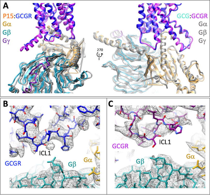
Figure 8.
Distinctions in the conformation of ICL1 between the P15-bound and GCG-bound GCGR structures alter the orientation of the Gs protein interface. A, ribbon representation of the protein backbone of GCGR and G protein subunits. GCGR in the P15 bound complex is blue and in the GCG bound complex, purple. In the P15 complex structure, Gαs is gold; Gβ1, cyan; Gγ2, dark purple. All subunits are colored gray in the GCG-bound complex. B, EM density map (contour 0.01) to model for GCGR ICL1 in the P15-bound complex. GCGR, blue; Gαs, gold; Gβ1, cyan. C, EM density map (contour 0.037) to model for GCGR ICL1 in the GCG bound complex. GCGR, purple; Gαs, gold; Gβ1, cyan.

