
Figure 5.
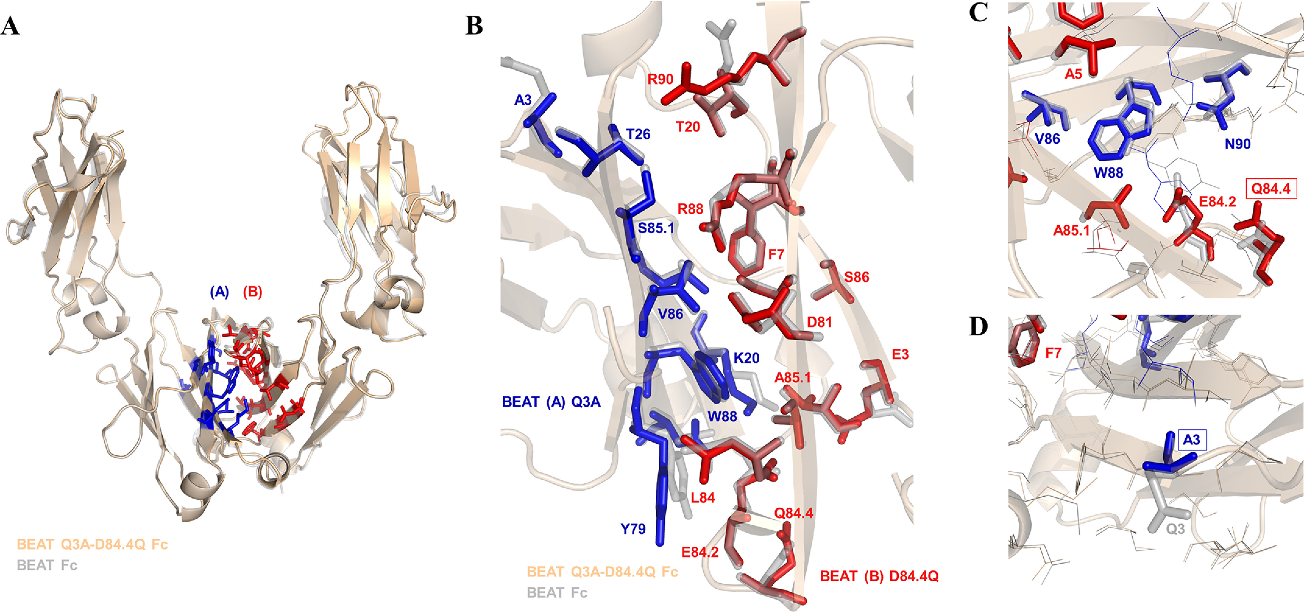
Crystal structure of the BEAT Q3A-D84.4Q Fc. A, ribbon diagram. Structural alignment of the original BEAT Fc structure (PDB code 5M3V) with that of BEAT Q3A-D84Q Fc (PDB code 6G1E). BEAT substitutions are in blue and red. B, close-up view of the same structural alignment. Side chains of the original BEAT Fc are in gray. A significant conservation of side-chain conformations could be observed (IMGT numbering). C, close-up showing position Gln84.4 and residues in its immediate environment, highlighting that no significant structural changes were induced by the mutation. D, close-up showing position A3 and residues in its immediate surroundings.