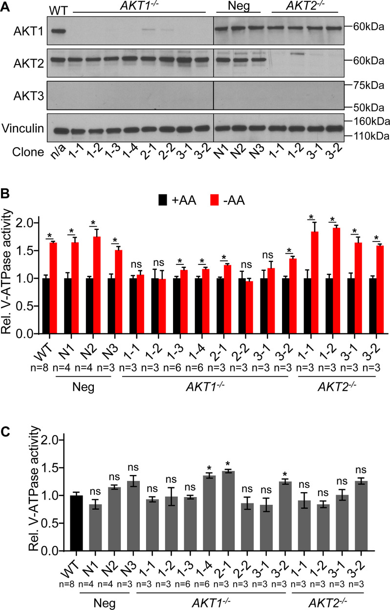
Figure 5.
AKT1 knockout HEK293T cells have reduced ability to increase lysosomal V-ATPase activity in response to amino acid starvation. A, 3 different guide sequences were used to target either AKT1 or AKT2 for CRISPR-mediated disruption in HEK293T cells. Western blotting was performed on lysates from untransfected WT cells, clones transfected with nontargeting control plasmids (Neg), clones targeted for disruption of AKT1 (AKT1−/−), and clones targeted for disruption of AKT2 (AKT2−/−). Isoform-specific AKT antibodies were used to assess protein knockdown, and vinculin was used as a loading control. Representative images are shown (n = 3). B, WT, nontargeted negative control (Neg), AKT1 knockout (AKT1−/−), and AKT2 knockout (AKT2−/−) HEK293T cells were incubated with FITC-dextran and then either maintained in amino acids (+AA) or starved for 1 h (−AA). The rate of V-ATPase–dependent fluorescence quenching in lysosomes was determined as described previously. Values are expressed relative to the unstarved condition for each clone tested. AKT2 knockout cells displayed a statistically significant increase in V-ATPase activity following amino acid starvation, whereas AKT1 knockout cells did not. *, p < 0.05; ns, p ≥ 0.05. Error bars represent S.E. C, Lysosomal V-ATPase activity for unstarved CRISPR negative control (neg), AKT1 knockout (AKT1−/−), and AKT2 knockout (AKT2−/−) cells is shown relative to unstarved WT cells. *, p < 0.05; ns, p ≥ 0.05. Error bars represent S.E.

