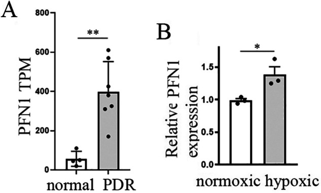
Figure 1.

PFN1's association with PDR and abnormal retinal angiogenesis. A, Transcripts per kilobase million (TPM) values for PFN1 in CD31+ ECs from diabetic (n = 7) versus control (n = 4) human retinal samples (based on GEO data set GSE94019) were calculated using Salmon and DESeq2 (p = 1.98E-11; FDR = 8.64E-9; log2FC = 3.35; data are the means ± S.D.). B, quantitative RT-PCR analyses of retinal PFN1 expression relative to the arithmetic mean of three housekeeping genes (GAPDH, Rps26, and Actb) in newborn mice relative to control mice at P17 (data are the means ± S.E. of 3 replicates/mouse; n = 3 mice/group).
