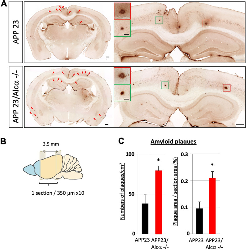
Figure 3.
Quantification of amyloid plaques in APP23 mouse brain in the presence or absence of Alcα. A, immunostaining of coronal sections of brain regions including the cerebral cortex and hippocampus of APP23 (top) and APP23/Alcα-deficient mice (bottom) at 12 months of age. The brain sections were stained with anti-human Aβ antibody. Arrowheads, typical amyloid plaques. A magnified view of the area around the hippocampus and cortex is shown on the right with magnified images of plaques (squares in red and green). Scale bar, 300 μm (sections) or 50 μm (plaques). B and C, 10 35-µm-thick sections with 315-µm intervals were examined in one mouse. The total plaque numbers in 10 sections/mouse were counted and are indicated as the number (left) or area (right) of plaques per area (left) or section area (right). Error bars, S.E. (unpaired t test; *, p < 0.05; 4 mice for APP23, 3 mice for APP23/Alcα-deficient background).

