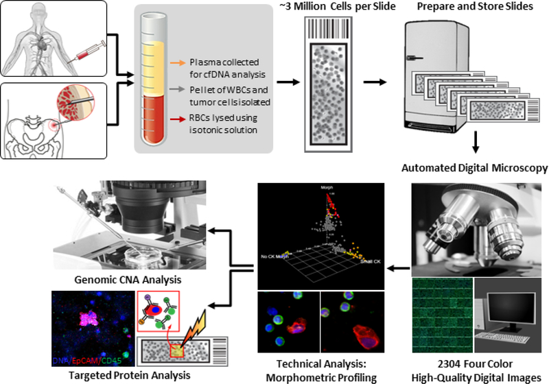
Figure 1.
HD-SCA platform for morphoproteogenomic profiling of liquid biopsy. PB and BMA samples are initially spun down for plasma extraction. Next, they undergo red blood cell lysis before plating approximately 3 million nucleated cells on each slide. Prepared slides are stored at −80 °C until needed for fluorescent antibody staining. Stained slides are first morphometrically profiled using automated digital microscopy at 10 × magnification, followed by classification by a technical analyst. Identified tumor cells are then re-imaged at 40 × magnification and proceed for genomic CNA or targeted protein analysis via imaging mass cytometry.

