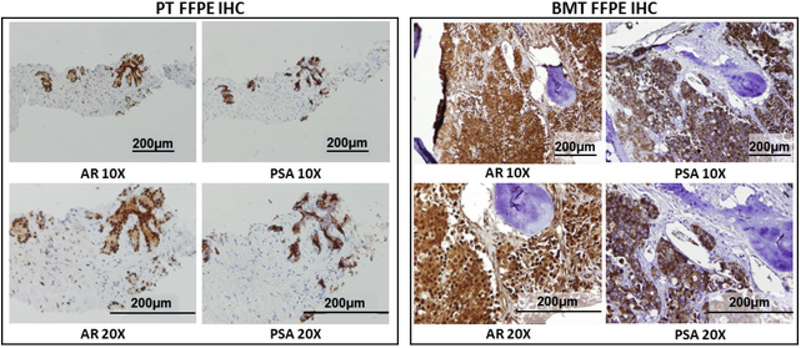
Figure 3.
IHC staining of FFPE PT from diagnostic PNBX and BMT from bone marrow biopsy. IHC staining for AR and PSA were performed on both PT and BMT samples. Samples were imaged using a light microscope at 10 and 20× objective. Staining results were reported as positive or negative for each marker.

