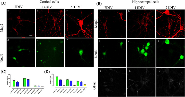
Figure 2. Cultured cortical and hippocampal primary neurons stained at three different time points for neuronal markers Map2, NeuN and GFAP.
(A) (a–f) Cortical neurons at 7 DIV (a,d), 14 DIV (b,e) and 21 DIV (c,f). (B) Hippocampal neurons at 7 DIV (a,d,g), 14 DIV (b,e,h) and 21 DIV (c,f,i). (C) Cell counting in cortical neurons using markers for neurons (NeuN), astrocytes (GFAP) and a dendritic cell marker, Map2. (D) Cell counting of the representative markers in hippocampal neurons. The neurons are stained with NeuN (green), Map2 (red) and GFAP (gray). Scale bar = 10 µm. Data represented as mean ± SEM. Abbreviations: DIV, days in vitro.

