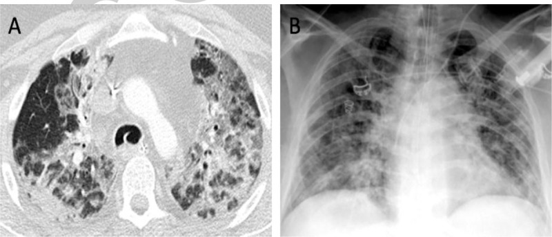
Fig. 2.
A pulmonary presentation of SARS-CoV-2 infection in a severely ill, intubated, and mechanically ventilated COVID-19 patient by computed tomography (CT; panel A) and plain chest x-ray imaging (panel B).

The CT shows characteristic milk-glass like opacities with consolidations in both upper lobes (A). CT findings may be unspecific and the primary diagnosis of SARS-CoV-2 remains laboratory-based. However, if indicated, imaging studies are helpful in assessing the severity and the course of COVID-19 pneumonia. A CT score can be used to evaluate the severity of the disease (15). The risks of an in-hospital transfer and potential contamination need to be considered. (Source: Axel Gossmann (MD), Department of Radiology, Cologne-Merheim Medical Center (CMMC), Cologne, Germany). COVID-19 indicates coronavirus disease 2019; SARS-CoV-2, severe acute respiratory syndrome coronavirus 2.
