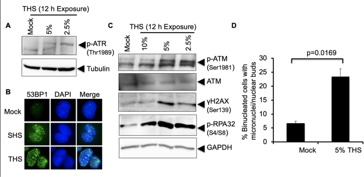
Fig. 4.

THS exposure causes replication stress and activates DSB response machineries. (A) Replication stress response was monitored following exposure of hPFs to the indicated doses of THS for 12 h followed by western analysis with anti-phosphoATR Thr1989 antibody. Tubulin was used as loading control. (B) Induction of DSBs by exposure of hPFs to 2.5% THS for 24 h was analyzed by IF detection of 53BP1 protein, which accumulates in foci at DSBs. SHS (0.4 PE) treatment was used as a positive control. (C) Activation of ATM following exposure of hPFs to 10%, 5% and 2.5% THS for 12 h was analyzed by western with anti-pATM (Ser1981). The same membrane was probed for phosphorylation of histone H2AX using anti-γH2AX antibody (Ser139) as an indicator of DSB formation and for pRPA32 (as in Fig. 3) to monitor replication stress. (D) Micronuclei formation in BEAS-2B cells following exposure to 5% THS for 48 h. Data represent the mean of ±SD for N=2.
