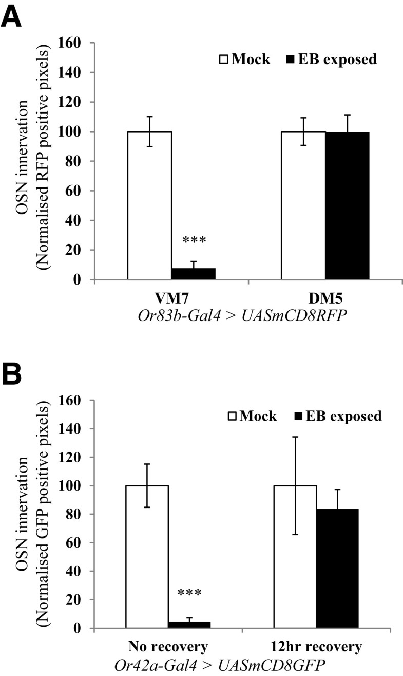
Figure 2.
Quantification of OSN innervation in antennal lobe glomeruli. A, OSN innervation after 2 d of EB exposure. OSNs that innervate various olfactory glomeruli were visualized in Or83b-Gal4> UAS-mCD8 RFP flies, using immunohistochemistry and confocal microscopy. Bar graph shows normalized RFP-positive pixels in VM7 and DM5 glomeruli of the same animals. B, Innervation of Or42a-positive OSNs is rapidly restored within 12 h after recovery after 4 d of odorant exposure. Or42a-Gal4 > UAS-mCD8GFP flies were exposed to EB for 4 d starting 0–12 h after eclosion, and then one group was allowed 12 h of recovery in air before dissection. Bar graphs show the extent of OSN innervation in VM7 glomeruli with and without 12 h of recovery. Error bars indicate the mean ± SEM. *p ≤ 0.05, **p ≤ 0.01, ***p ≤ 0.001, determined by Student's t test.

