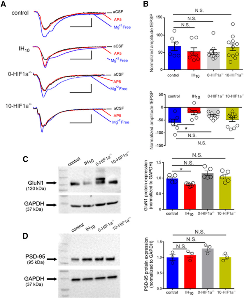
Figure 3.
The IH reduces the contribution of the NMDAr to fEPSP and GluN1 protein from wild-type mice but does not induce these changes in HIF1a+/−. A, Representative traces of the fEPSP from control, IH10, 0-HIF1a+/−, and 10-HIF1a+/− in: aCSF (black), Mg2+-free media (blue), and Mg2+-free media with AP5 (red). Scale bars: 0.4 mV/10 ms. B, top, Change in amplitude of the fEPSP from aCSF to Mg2+-free media. Bottom, Change in amplitude of the fEPSP from Mg2+-free media to Mg2+-free media with AP5; *p < 0.05; N.S., p > 0.05. C, left, Representative Western blottings of GluN1 and the housekeeping protein, GAPDH from control (n = 5), IH10 (n = 5), 0-HIF1a+/− (n = 5), and 10-HIF1a+/− (n = 5). Right, Comparisons of normalized GluN1 protein expression were performed to compare experimental conditions to control. This revealed that GluN1 was reduced in IH10 and unchanged in both 0-HIF1a+/− and 10-HIF1a+/−; *p < 0.05; N.S., p > 0.05. D, left, Representative Western blottings of PSD-95 and the housekeeping protein, GAPDH from control (n = 3), IH10 (n = 3), 0-HIF1a+/− (n = 3), and 10-HIF1a+/− (n = 3). Right, Comparisons of normalized PSD-95 protein expression were performed to compare experimental conditions to control; *p < 0.05; N.S., p > 0.05.

