Figure 5.

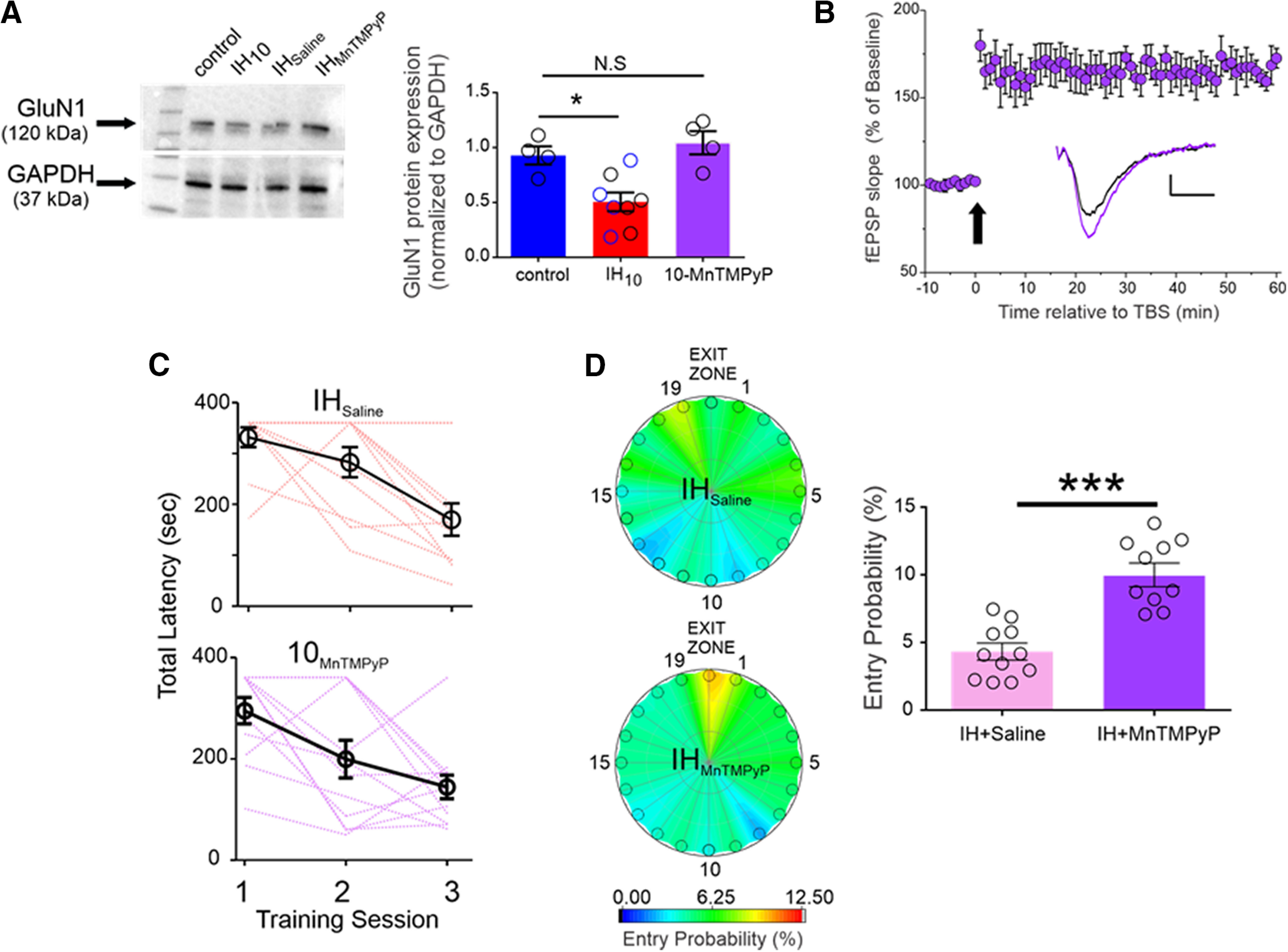
Antioxidant treatment mitigates the IH-dependent effects on GluN1 expression, LTPTBS, and performance in the Barnes maze. A, left, Representative Western blottings of GluN1 and GAPDH from Control, IH10, wild-type mice treated with saline during 10 d of IH (i.e., vehicle control exposed to IH, IHSaline, n = 4), wild-type mice treated with MnTMPyP during 10 d of IH (IHMnTMPyP). Right, Normalized GluN1 protein expression was examined in control (n = 4), IH10 (n = 4), IHSaline (n = 4), IHMnTMPyP (n = 4). No difference in GluN1 was evident between IH10 (open black circles in IH10 label) and IHSaline (open blue circles in IH10 label); therefore, the two groups were merged into the IH10 label for comparisons to control. Comparisons revealed that GluN1 was reduced only in IH10 and unchanged in IHMnTMPyP. B, In hippocampal slices from IHMnTMPyP, LTPTBS (n = 5) could be reliably evoked contrasting the effect of IH10 on LTPTBS (Fig. 2B). Scale bars: 0.2 mV/10 ms. The arrow represents the TBS protocol. C, The total latency to exit the Barnes maze progressively decreased in both IHSaline (n = 11, pink lines represent individual performance) and IHMnTMPyP (n = 10, purple lines represent individual performance), suggesting that both groups could learn the exit zone location. D, Heat maps of the mean entry probability across all false exits (1–19) and the exit zone during the probe trial for IHSaline and IHMnTMPyP. Comparison of entry probability into the exit zone during the probe trial reveals that IHMnTMPyP has a greater probability for entering the exit zone when compared with IHSaline (p = 0.006); ***p < 0.001, *p < 0.05; N.S., p > 0.05.
