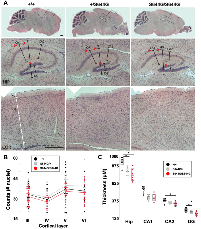
Figure 3.
Aberrant hippocampal morphology of Grin2aS644G adolescent mice. (A) Haematoxylin and eosin stained brain sections (scale bars = 500 µm). At postnatal Day 14, markedly decreased hippocampal thickness, particularly in CA1 and DG (arrowheads). (B) Cerebral cortex, showing no consistent significant differences in width between genotypes, proxied by cell counts per layer after accommodating random variation between replicates. (C) Decreased thickness in the hippocampus, particularly in CA1 and DG. n = 3 for each genotype, counts or thickness measured from both hemispheres. Error bars are quantiles. *P < 0.05; Steel-Dwass non-parametric means comparison.

