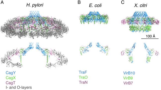
Fig. 4.
Structural comparison of the H. pylori Cag T4SS OMCC with minimized T4SSs from other bacterial species. (A-C) Upper panel, secondary structural models of the complexes. Lower panel, Central axial slice views of the secondary structural models. Scale bar, 100 Å. (A) OMCC from the H. pylori Cag T4SS (16). CagY (PDB-6ODI, blue), CagX (PDB-6OEG, green), CagT (PDB-6OEE, purple), and outer- and inner-layer proteins (PDB-6OEF and -6OEH, grey). (B) Conjugation system (encoded by pKM101) (66). TraF (PDB-3JQO, blue), TraO (PDB-3JQO, green), and TraN (PDB-3JQO, purple). (C) Xanthomonas citri T4SS (67). VirB10 (PDB-6GYB, blue), VirB9 (PDB-6GYB, green), and VirB7 (PDB-6GYB, purple).

