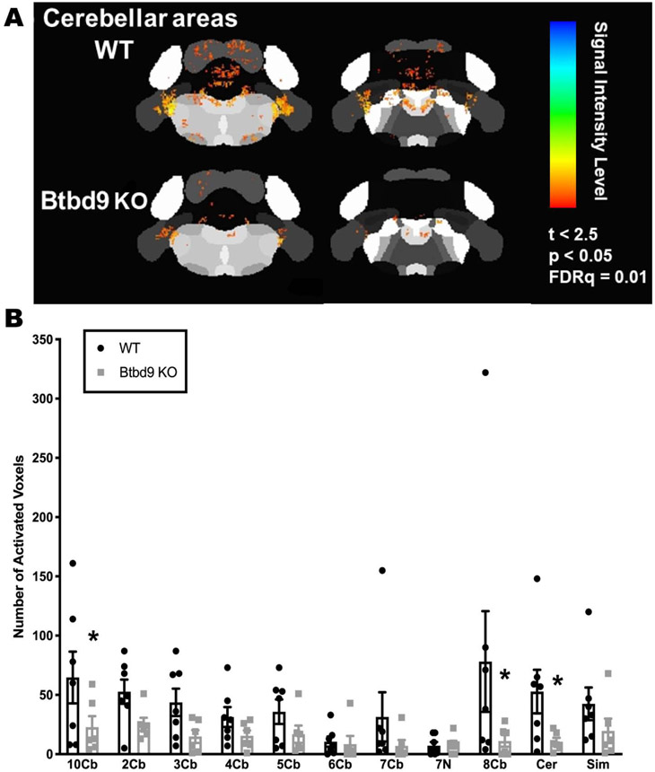
Figure 1.
MEMRI recordings of neural activity in cerebellar regions of the systematic Btbd9 KO mice (n=6) and their WT littermates (n=8). Btbd9 KO mice had significantly decreased neural activity in cerebellar lobules VIII, cerebellar lobules X and deep cerebellar nuclei. Cb: cerebellum lobules; 7N: facial nucleus; Cer: cerebellar nuclear area; Sim: Simple lobule. Bars represent means plus standard error of means (SEMs). *, p ≤ 0.05.

