Figure 4.
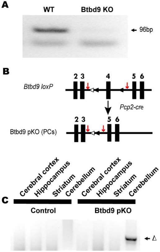
Generation and molecular characterization of Btbd9 pKO mice. (A) No Btbd9 mRNA in the cerebellum of the Btbd9 KO mice. (B) Schematic diagram of the generation of the Btbd9 pKO mice. Filled boxes represent exons. Filled triangles indicate loxP sites. Open triangles indicate the FRT sites that were incorporated to remove the neo cassette. In Btbd9 pKO mice, exon 4 is deleted in PCs because cre is expressed specifically and the recombination occurs in the PCs. The red arrows indicate the sites of the PCR primers used to detect the recombination event. (C) Tissue-specific deletion of Btbd9 exon 4 in Btbd9 pKO mice was confirmed by PCR using DNA isolated from each brain region. The deletion (Δ) was detected only in the cerebellum (includes PCs) of Btbd9 pKO mice as predicted.

