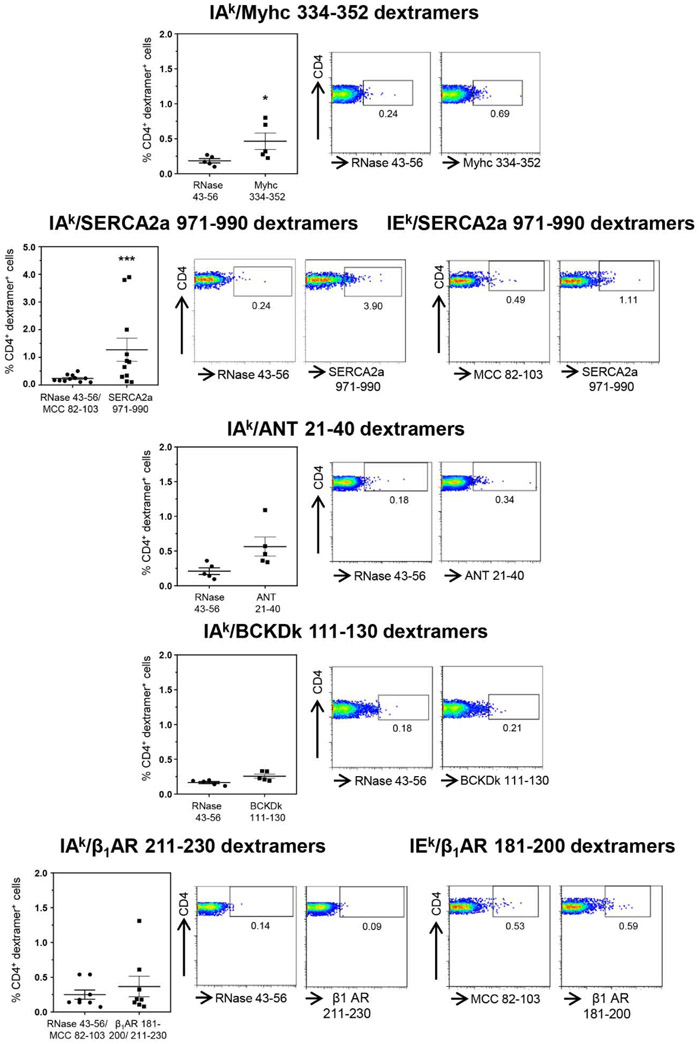
Figure 1: Detection of autoreactive T cells with multiple antigen-specificities in animals infected with CVB3.
Lymphocytes obtained from animals infected with CVB3 were stimulated with Myhc 334-352, SERCA2a 971-990, ANT 21-40, BCKDk 111-130, and β1AR 181-200 or β1AR 211-230 for three days, and cells were rested in IL-2 medium. Cells harvested on day 8 to 10 post-stimulation were stained with the indicated IAk and/or IEk dextramers, anti-CD4 and 7-AAD. After washing, and acquiring by flow cytometry, dext+ CD4+ cells were enumerated. Mean ± SEM values obtained from three to six individual experiments, each involving three to six mice are indicated in the left panel for each dextramer reagent. Representative flow cytometric plots for control and specific dextramers are shown on the right. RNase 43-56, control for IAk dextramers; and MCC 82-103, control for IEk dextramers. *P < 0.05 and ***P < 0.001.

