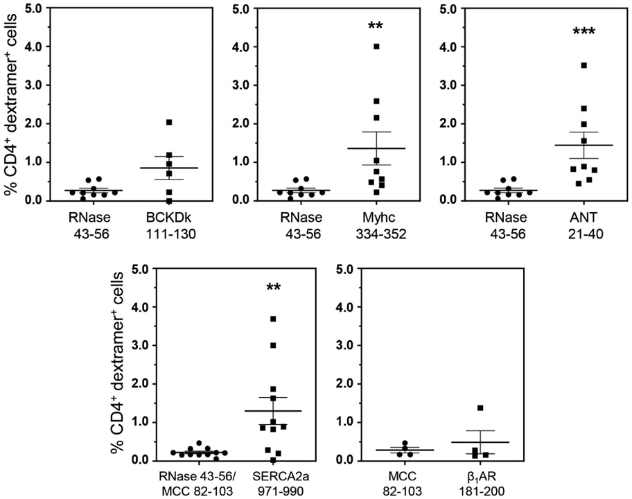
Figure 2: Detection of autoreactive T cells in the livers from CVB3-infected mice.
Livers from CVB3 infected mice were processed to obtain MNCs as described in the methods section. MNCs were stained with the indicated IAk and/or IEk dextramers, followed by anti-CD4, and 7-AAD. After acquiring the cells by flow cytometry, dext+ cells were analyzed in the live (7-AAD−), CD4 subset. Mean ± SEM values from four to nine individual experiments, each involving three to six mice are shown. RNase 43-56 (control for IAk dextramers); and MCC 82-103 (control for IEk dextramers). **P < 0.01, and ***P < 0.001.

