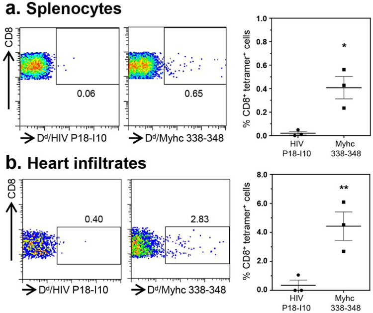
Figure 4: CVB3-infection leads to the generation of Myhc 338-348 tet+ CD8+ T cells.
(a) Splenocytes. Between 18 to 21 days post-infection with CVB3, splenocytes were prepared and stimulated with Myhc 334-352 for three days. After resting in IL-2 medium, cells harvested on day 8 or 9 were stained with CD4 and CD8 antibodies and 7-AAD and H-2Dd/Myhc 338-348 (specific) and HIV P18-I10 (control) tetramers. Cells were washed and acquired by flow cytometry to determine the frequencies of tet+ cells in the live (7-AAD−) CD8 subset. (b) Heart infiltrates. Hearts collected from CVB3-infected animals were processed to obtain MNCs. Cells were stained as above, and after acquiring by flow cytometry, frequencies of live (7-AAD−), tet+ CD8+ cells were enumerated. Representative flow cytometric plots and mean ± SEM values derived from four experiments each involving two to three mice are shown. *P<0.05 and **P<0.01.

