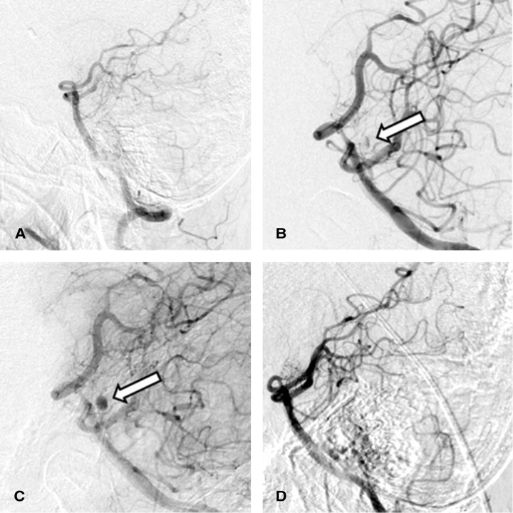
Fig. 2.
(A) Initial DSA was negative for the source of hemorrhage. (B) DSA performed on day 39 showed a 3 mm aneurysm (arrow) at the posterior surface of the upper third of the basilar artery. (C) The aneurysm (arrow) was observed in the late arterial phase of the same DSA, as shown in (B), without directly involving the basilar trunk. (D) DSA performed on day 64 showed complete resolution of the aneurysm. DSA: digital subtraction angiography.

