Figure 1.
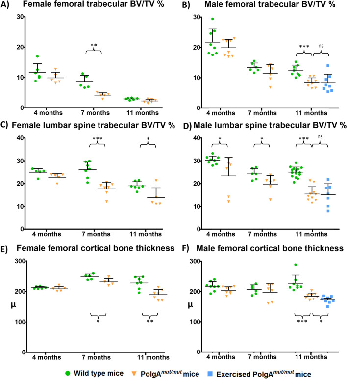
Micro-CT scan of femurs and lumbar spines demonstrate accelerated bone loss in PolgAmut/mut mice. An exercise intervention was not associated with a change in trabecular bone mass. A significant decline in femoral trabecular BV/TV can be seen by 7 months of age in female (A) PolgAmut/mut mice (p = 0.003) and by 11 months of age in male (B) PolgAmut/mut mice (p < 0.0001) when compared to age matched wild type controls (unpaired 2 tailed t-test). A similar pattern of bone loss was seen in the lumbar spine with significant reductions in trabecular BV/TV in female PolgAmut/mut mice at 7 and 11 months (C), and in male PolgAmut/mut mice at 4, 7 and 11 months (D), compared to age and sex matched wild type controls. Cortical thickness was also significantly reduced in female PolgAmut/mut mice at 7 and 11 months (E), and in male PolgAmut/mut mice at 11 months (F), compared to wild type controls. Exercise had no significant effects (ns) on bone mass observed in 11 month old PolgAmut/mut males in terms of trabecular BV/TV (B, D). A gradual decline in BV/TV levels is observed in wild type mice as a feature of advancing age, with females showing a significantly reduced femoral trabecular BV/TV in comparison to age matched males at 4, 7 and 11 months of age (p = 0.0004, p = 0.0015, p < 0.0001 respectively (unpaired 2 tailed t-test)).

