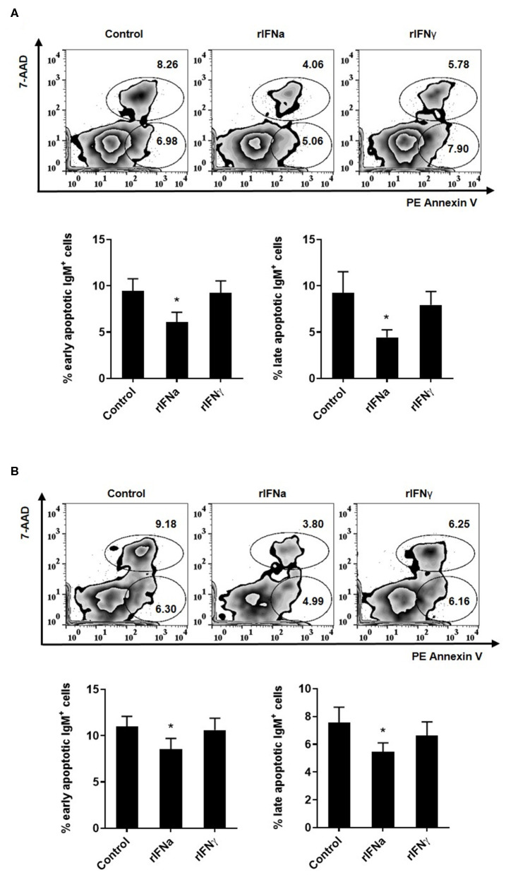
Figure 3.
Effect of type I and type II IFNs on the spontaneous apoptosis of blood IgM+IgD+ B cells. PBLs were incubated for 48 h (A) or 72 h (B) at 20°C with 50 ng/ml rIFNa, 20 ng/ml rIFNγ or media alone (control). Then, cells were stained with anti-trout IgM for 20 min at 4°C. After washing, PE Annexin V and 7-AAD were used to stain the cells and identify early (Annexin V+/7-AAD−) and late (Annexin V+/7-AAD+) apoptosis. Data were analyzed within 1 h as described in the Materials and Methods section. Representative dot plots are presented along with graphs showing the quantification of apoptotic IgM+ B cells (mean + SEM; n = 6–9). Asterisks denote significant differences between samples treated with rIFNs and control samples (*P ≤ 0.05).

