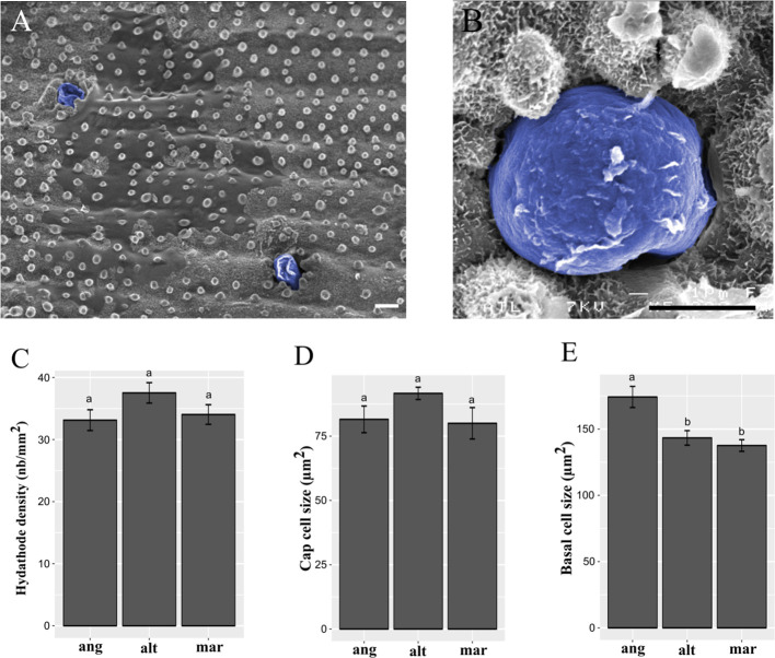
Figure 3.
Spartina salt gland structure and observation. Salt glands were observed in ridges of the adaxial leaf surface by scanning electron microcopy, and colored in blue after taking photographs (A, B). Scale bars = 5 µm. We measured salt gland densities (C; in gland.mm-2), cap cell size (D; in µm2), and basal cell size (E; in µm2) in the allododecaploid Spartina anglica (ang) and its related parents S. alterniflora (alt) and S. maritima (mar). Values annotated with different letters between species are significantly different according to Kruskal-Wallis’s multiple comparison test (with Bonferroni correction, p.value < 0.05).

