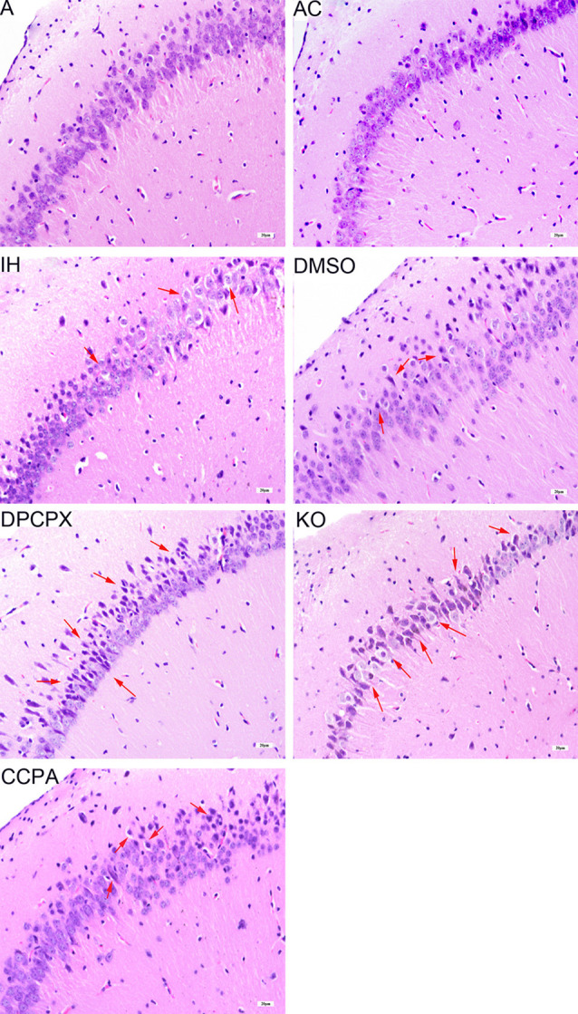
Figure 2.

Neuronal morphology in the hippocampal CA3–CA1 region detected by hematoxylin and eosin staining. Arrows indicate swollen and heterologous neurons with clear nuclei. Bar: 20 μm. A, room air; AC, intermittent air; IH, intermittent hypoxia; DMSO, IH with DMSO treatment; CCPA, IH with 2-chloro-N (6)-cyclopentyl-adenosine treatment; DPCPX, IH with 8-cyclopentyl-1,3-dipropylxanthine treatment; KO, IH with A1R knockout.
