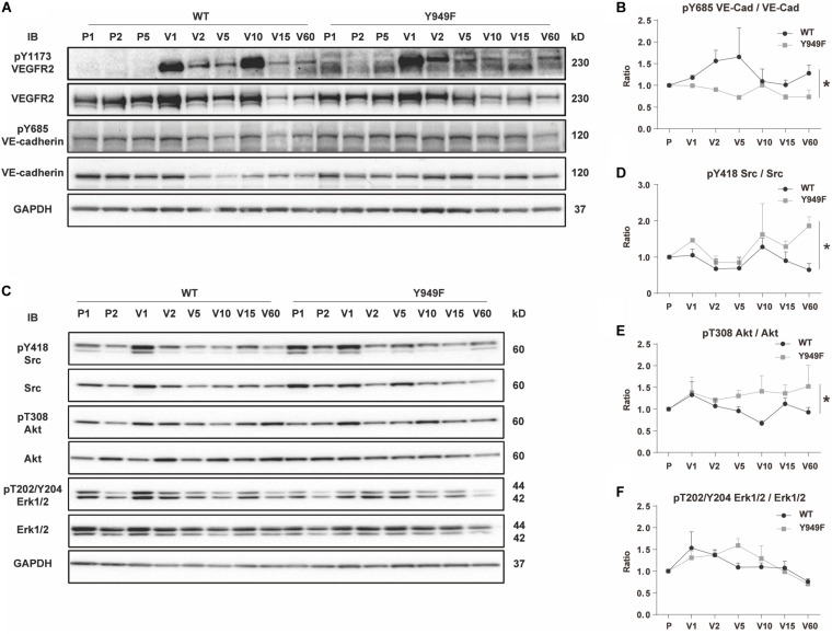
FIGURE 4.
VEGF-induced VE-cadherin pY685 accumulation in hearts. (A) Immunoblot for pY1173 VEGFR2 and total VEGFR2, and pY685 VE-cadherin and total VE-cadherin on heart lysates from WT and Vegfr2Y949F/Y949F mice. Mice were injected with PBS or VEGFA in the tail-vein, followed by circulation for different time periods; PBS was injected and left to circulate for 1, 2 or 5 min (denoted P1, P2, P5). Alternatively VEGFA was injected and left to circulate for 1, 2, 5 min etc. (denoted V1, V2, V5 etc.). Heart lysates were immunoblotted for 3-phosphate dehydrogenase (GAPDH) as a loading control. (B) Quantification of pY685 VE-cadherin normalized to total VE-cadherin in samples shown in (A). (C) Immunoblotting for pY418 c-Src and total c-Src, pT308 Akt and total Akt, pT202/Y204 Erk1/2 and total Erk1/2. (D–F) Quantification of phosphorylated protein bands normalized to the corresponding total protein. n = 3; i.e., three independent replicates of experiments as shown in panel (A) with one mouse/time point for each strain and nine mice in total/strain for each experiment. Wilcoxon test, *p < 0.05.

