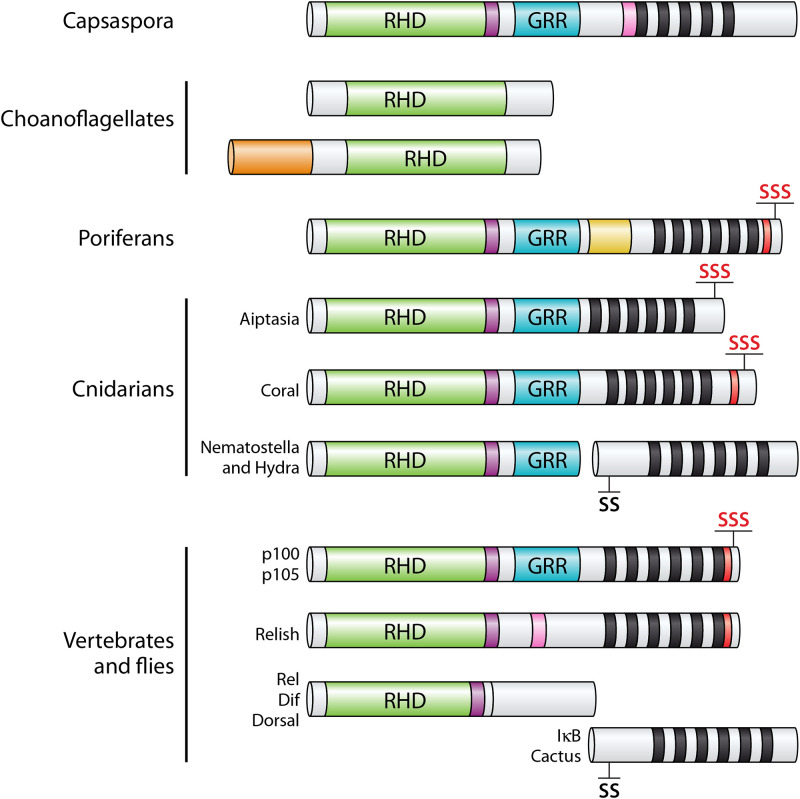
FIG 1.
Structures of NF-κBs in organisms basal to Bilateria are most similar to human NF-κB proteins rather than Rel proteins. The basic domain structures of homologs of NF-κB in Capsaspora, choanoflagellates, poriferans (sponges), and cnidarians are compared to those of the fly and vertebrate NF-κBs (Relish and p100/p105), Rels (Dif, Dorsal, RelA, RelB, and c-Rel), and IκB (Cactus). Green, Rel homology domain (RHD); purple, nuclear localization signal (NLS); blue, glycine-rich region (GRR); pink, caspase cleavage site; black bars, ankyrin repeats; red, death domain; SSS/SS, conserved serines important for phosphorylation and degradation of the protein (the red SSSs indicate homology to p100); orange, N-terminal domains of choanoflagellates not seen in other NF-κB proteins.

