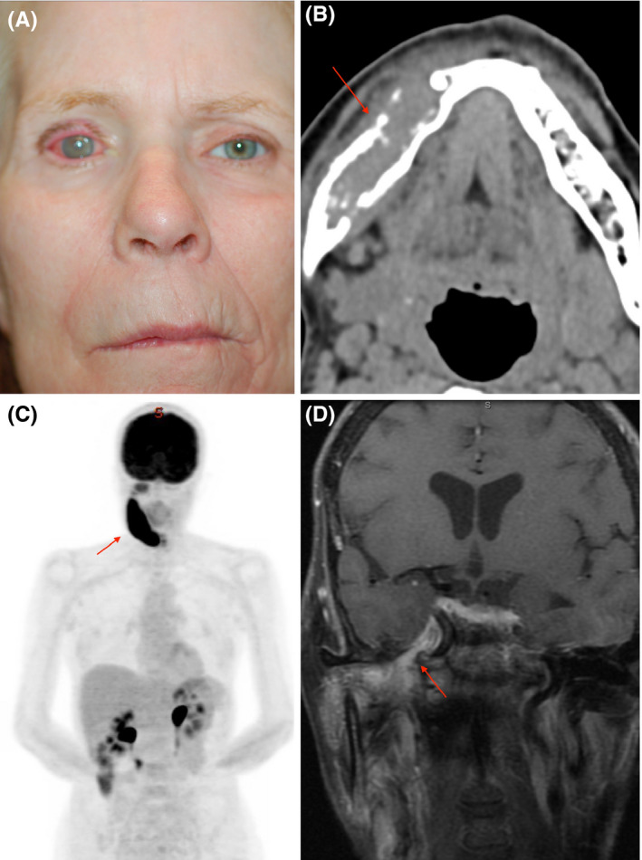
Figure 2.

A, External photograph showing right keratopathy, facial nerve paralysis, and temporal wasting (B). Axial computed tomography (CT) demonstrating an osseous lesion of the right mandible (red arrow). C, Fluorodeoxyglucose (FDG)‐positron emission tomography (PET) with FDG uptake of the right mandible (red arrow). D, MR coronal T1 FSGD with enhancement of the trigeminal nerve (red arrow)
