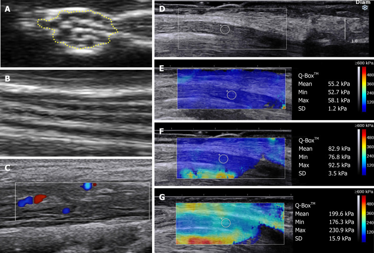
Figure 1.
Approaches and techniques used in neuromuscular ultrasound. A and B: Normal nerve ultrasound appearances of the median nerve in the forearm in B-mode. A: Cross-sectional area traced within the hyperechoic rim; B: Longitudinal view demonstrating the normal linear fascicular pattern; C: Doppler signal in the tibial nerve of a patient with hypertrophic perineuritis; D-G: Sheer wave elastography in carpal tunnel syndrome; D: Longitudinal B-mode image of the median nerve at the carpal tunnel above the lunate bone, a typical location for elastography measurements; E: Elastography values in a normal wrist; F: increased median nerve stiffness in mild carpal tunnel syndrome; G: further increased median nerve stiffness in severe carpal tunnel syndrome. D-G: Citation: Wee TC, Simon NG. Ultrasound elastography for the evaluation of peripheral nerves: A systematic review. Muscle Nerve 2019; 60: 501-512. Copyright© The Authors 2020. Published by John Wiley and Sons.

