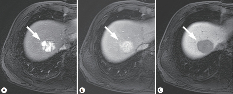
Figure 2.
HCC in a 53-year-old man with chronic hepatitis B. On the arterial (A), portal venous (B), and hepatobiliary phase (C) images, after administration of hepatobiliary agent, a 39-mm liver mass (arrows) showed arterial phase hyperenhancement without washout in the portal venous phase, while showing hypointensity in the hepatobiliary phase. The mass was categorized as LR-4 by LI-RADS 2018, but classified as definite HCC by KLCA-NCC 2018. HCC, hepatocellular carcinoma; LI-RADS, Liver Imaging Reporting and Data System; KLCA-NCC, Korean Liver Cancer Association-National Cancer Center.

