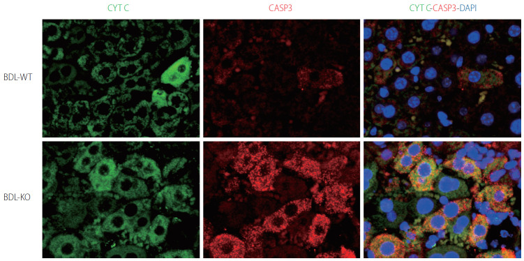
Figure 5.
Hepatocyte apoptosis in Cygb-KO liver under BDL. Expression of markers of apoptosis cytochrome C (CYTC) (green) and active + pro Caspase (CASP) 3 (red) after 24 hours of BDL in WT and KO mice. DAPI, blue, was used as nuclear counterstain. Original magnification, ×1,200. BDL, bile duct ligation; Cygb, cytoglobin; DAPI, 4’,6-diamidino-2-phenylindole.

