
Figure 1.
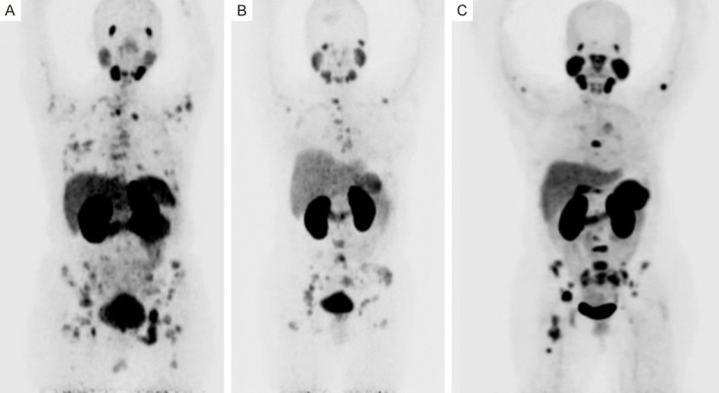
A. Maximal intensity projection (MIP) Positron Emission Tomography PET with 68Ga-PSMA in patient with TPN breast cancer, showed normal biodistribution and primary site in right breast, multiple bone and lymph-node metastases. B. MIP in patient with LUM-B HER2 positive breast cancer, showed multiple bone metastases with faint uptake in primary site in the left breast. C. MIP in patient with Her-2 overexpression breast cancer, showed multiple bone metastases with faint uptake in primary site in the right breast.