
Figure 2.
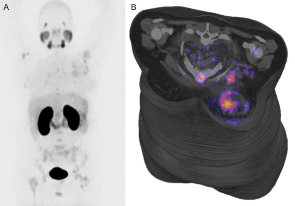
(A) Maximal intensity projection (MIP) Positron Emission Tomography PET with 68Ga-PSMA in patient with LUM-A breast cancer, showed normal biodistribution and primary site in left breast, multiple bone and axillar lymph-node metastases are present; in the left breast are present necrosis in center of tumor (B).