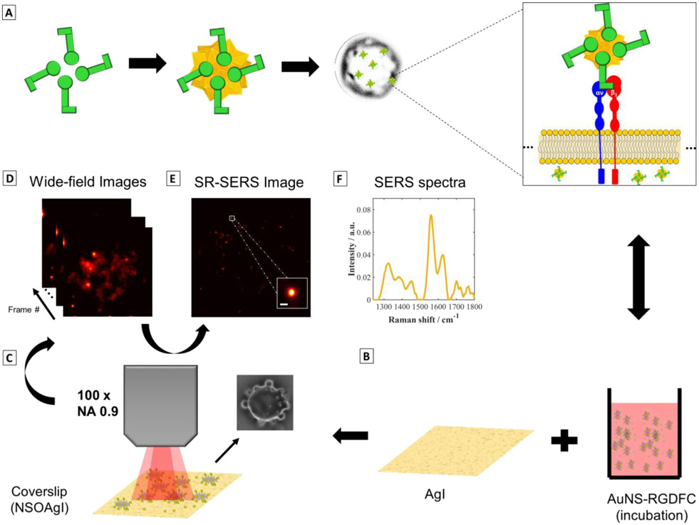
Figure 1. Super-resolution SERS imaging of AuNS-RGDFC in cancer cells.
(A) RGDFC-functionalized AuNS were used as label free SERS probes. AuNS had an average size of 110±2nm and the LSPR peak max was at 657 nm (See supporting information, Figure S1 and Table S1). RGDFC functionalized AuNS were incubated with SW620 colon cancer cells in RPMI media to enable the AuNS-RGDFC to bind with αυβ3 integrin receptor as previous demonstrated. (B) The cells were fixed on Ag island covered coverslips. (C) Wide-field SERS images were collected using a 659 nm laser, with a power density of ~0.2 kW/cm2, and a 100× objective (NA 0.9) and 100 ms per frame. The inset image shows a bright-field image of a cell observed in this experiment. (D) A series of images recorded by collecting the Stokes scattering use an edge filter to reject the Rayleigh scattered light. (E) Super-resolution algorithms are applied to the wide field SERS images (separated in frames) to increase the localization of the AuNS probes interacting with the cell. The data shown was processed using ThunderSTORM50, with the following parameters adjusted for our camera: 65 nm per pixel, 1.6 photoelectrons per count, and camera base level of 50. The inset shows the improvement in the resolution when compared with the images in (D). The inset scale bar is 100 nm. (F) A typical Raman spectrum collected in this experiment is shown. The spectrum is obtained by directing the collected light through an optical fiber and then to a spectrograph and CCD. The integration time for the spectrum shown was 30 s.

