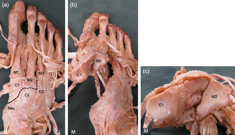
Fig. 1.
The procedure of the ligament dissection. a: Dorsal view of the right foot. b: Dorsal proximal view of the right foot. c: Articular surface of the first cuneiform and the second metatarsal in the Lisfranc joint. Black dotted line: the parts between navicular bone and the first cuneiform, between the second cuneiform and the second metatarsal, between the second metatarsal and the third metatarsal were separated. White dotted line: the parts between the first metatarsal and first cuneiform, between the first metatarsal and the second metatarsal were separated partially. 1: Lisfranc ligament, 2: cuneiform 1-metatarsal 2&3 plantar ligament, L: lateral, M: medial, C1: first cuneiform, C2: second cuneiform, C3: third cuneiform, M1: first metatarsal, M2: second metatarsal, M3: third metatarsal, Nav: navicular

