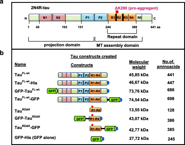
Fig. 2.
Tau proteins and variants used in this study. (a) Bar diagram of longest isoform of Tau in human CNS comprising 441 amino acids (Uniprot P10636-F, alias hTau40, Tau-2N4R). Domains are depicted with 2 near-N-terminal inserts (N1, N2, pink), the proline-rich domains (P1, P2; blue), 4 pseudo-repeats (R1-R4, ~ 31 residues each, ochre, plus one less-conserved repeat R5), and the C-terminal tail. The amyloidogenic hexapeptide motifs are indicated at the beginning of R2, R3 (thick black line). The C-terminal half (MT assembly domain P2-R5) promotes both microtubule assembly and pathological aggregation of Tau, the N-terminal half (projection domain) projects from the surface of microtubules or from the core of Tau fibers, respectively. The “pro-aggregant” deletion mutation ΔK280 (red dot) lies near the beginning of R2, it increases the β-propensity of Tau and hence increases pathological aggregation. (b) Tau and GFP fusion proteins are schematically presented with their name listed on the left. The GFP-tag is positioned either at the N-terminus or C-terminus. Some constructs contain a C-terminal 6xHis-tag to aid in the purification (blue ellipse). A sequence of 13–14 amino acids was used as a linker (black line) in the two short Tau constructs GFP-TauRDΔK and TauRDΔK-GFP to increase the distance between the GFP and the repeat domain (see Table S1)

