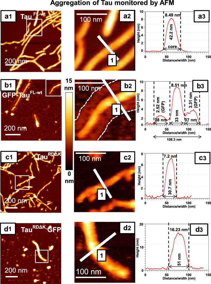
Fig. 5.
Atomic force microscopy reveals the changes in Tau filament assembly and decoration with GFP. AFM analysis of Tau fibrils formed in the presence of heparin for 24 h at 37 °C was imaged in tapping mode using an MSNL cantilever. Left row, overviews; center row, magnified details; right row, line scans showing distribution of height. TauFL-wt(a1) and GFP-TauFL-wt(b1) both form long fibrils (straight or twisted), but the height distribution shows characteristic differences because the attachment of GFP generates a halo on both sides of the filament core (compare a3 and b3, core and side bands indicated by dashed lines). The average height (thickness) of the fibril core is similar for TauFL-wt (7.7 nm ± 0.7 nm; n = 36) and GFP-TauFL-wt (7.0 nm ± 1.2 nm; n = 36). However, the overall thickness increases from TauFL-wt alone (38.1 nm ± 5.45 nm; n = 50) to GFP-TauFL-wt (96.01 nm ± 8.98 nm; n = 50). The two side peaks (b3) are ~ 3 nm wide, in good agreement with the shape of GFP. (c-d) The repeat domain constructs TauRDΔK forms well twisted filaments (c1) whereas TauRDΔK-GFP forms only few short filaments and more globular shaped aggregates (d1). The enlarged images (c2, d2) show that the short filaments of TauRDΔK-GFP are thicker than those of TauRDΔK. (c3 and d3) The widths and heights of TauRDΔK-GFP (width - 42.95 nm ± 8.32 nm; n = 50; height- 18.5 nm ± 3.1 nm; n = 36) fibrils are larger than those of TauRDΔK fibrils (width - 28.17 nm ± 3.35 nm; n = 50; height - 7.0 nm ± 1.1 nm; n = 36). The height scale for a1, b1, c1 and d1 is 0 to 15 nm

