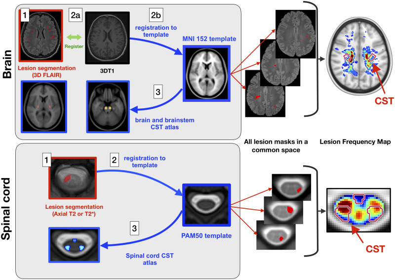
Figure 1.
Processing pipeline. Brain data processing: (1) semi-automatic lesion segmentation on 3D T2-FLAIR or axial T2-weighted images (when 3D T2-FLAIR was not available); (2a) intra-subject linear registration between T2-FLAIR and T1-weighted images; (2b) Affine and non-linear registration between T1-weighted images and the T1 ICBM 1 mm isotropic template space; and (3) quantification of lesion volume fraction based on brain and brainstem CST atlases. Spinal cord data processing: (1) manual lesion segmentation on axial T2*-weighted images; (2) slice-wise non-linear registration to the PAM50 template; and (3) Quantification of lesion volume fraction based on a spinal cord CST atlas. To create the lesion frequency maps, brain and spinal cord lesion masks were averaged in the ICBM and PAM50 space, respectively.

