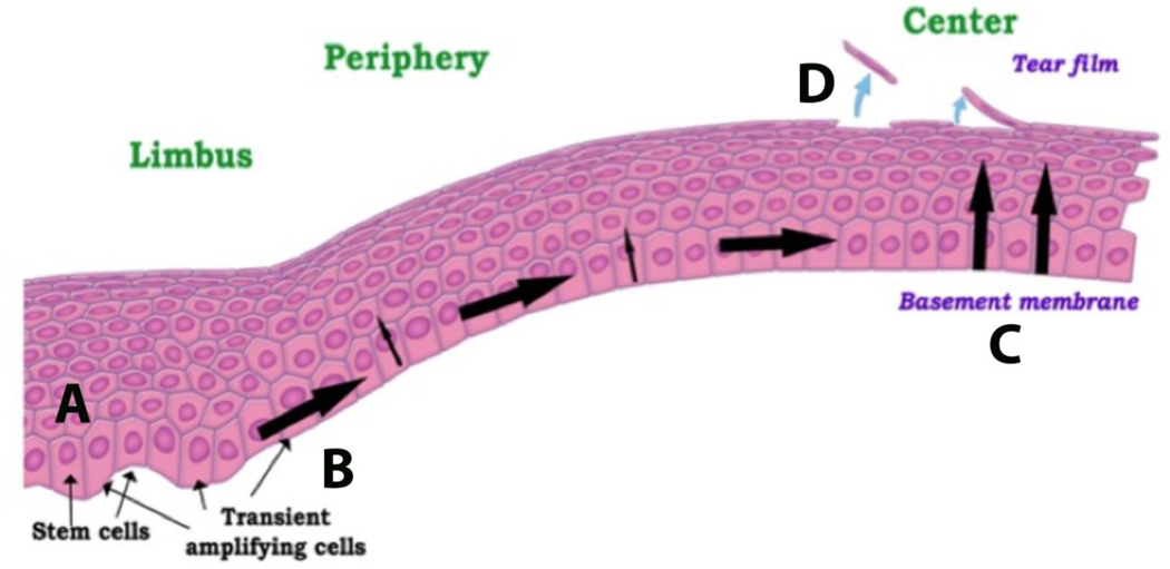
Figure 1:
Schematic of corneal epithelial renewal. (A) Stem cells reside in the basal layer of the limbus. (B) Following departure from the limbus, basal epithelial cells become transient amplifying cells and exhibit a high proliferative capacity. (C) Cells continue to migrate to the central cornea, losing proliferative capacity as they go. After the final round of cell division, the paired cells move towards the corneal surface. (D) At the corneal surface, cells are shed or desquamated into the precorneal tear film. Figure taken from Ladage et al. Contact Lens Ant eye 2002.

