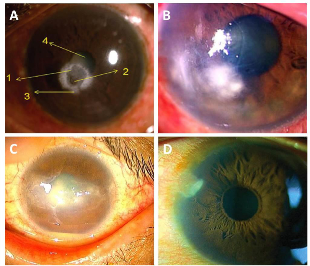
Figure 5:

Corneal ulcers reported in diabetic patients. (A) Corneal ulcer caused by Prototheca wickerhamii. Numbers as described as detailed in the original case report. 1: central ulcer; 2: region of corneal thinning; 3: large infiltrate surrounding the ulcer; 4: lenticular changes. Image taken from Narayanan et al. Indian J Ophthalmol 2018. (B) Corneal ulcer caused by Roussoella solani. Image taken from Mochizuki et al. J Infect Chemo 2017. (C) Corneal ulcer caused by Corynebacterium propinquum. Image taken from Todokoro et al. J Clin Microbiol 2015. (D) Corneal ulcer caused by Stenotrophomonas maltophilia. Image taken from Holifield et al. Eye Contact Lens 2011.
