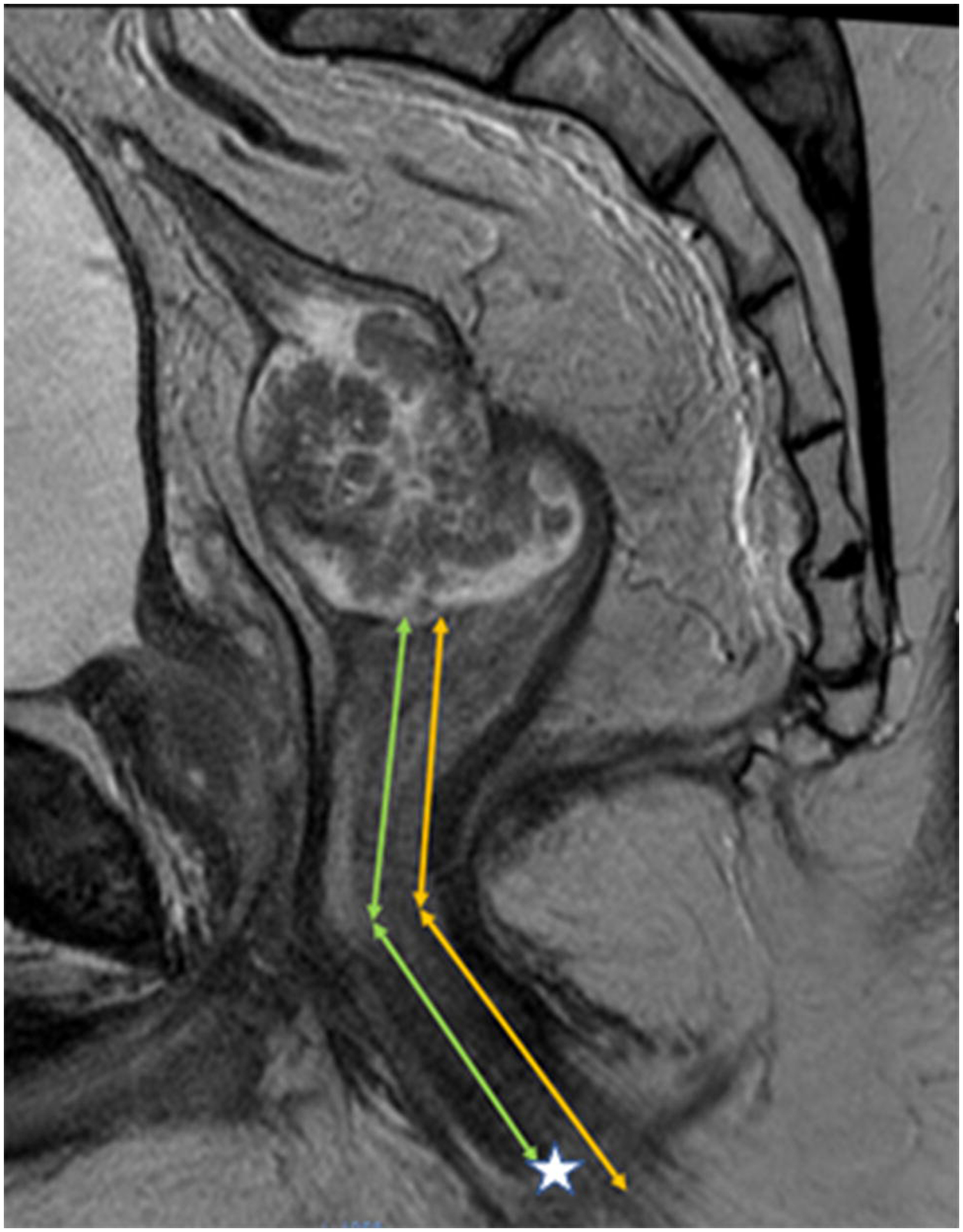
Figure 1.

Sagittal T2 weighted MRI of the pelvis in 42-year-old male with a polypoid rectal adenocarcinoma. Measurement of height of tumor from lower border of external anal sphincter [EAS] (corresponding to histologic anal verge), yellow lines. Measurement of height of tumor from bottom of internal anal sphincter [IAS] (often corresponding to surgical anal verge due to anesthesia induced skeletal muscle relaxation or forcible manual separation of EAS at time of sigmoidoscopy. Difference in these distances is only about 1-cm and represents the intersphincteric notch (asterisk).
