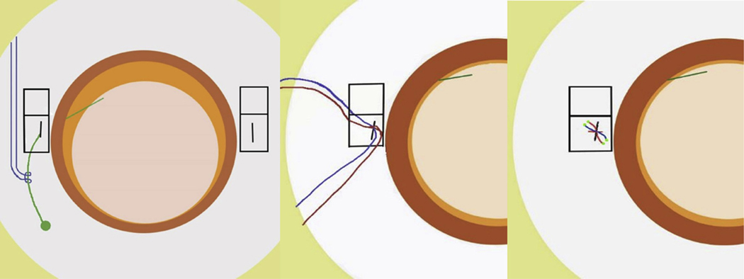
FIGURE.
Suture-tying technique used for 10–0 polypropylene fixation suture knot around the haptic of a posterior chamber intraocular lens. (Left) Haptic has been externalized through the fixation sclerotomy made 2 mm posterior to the surgical limbus. The 10–0 polypropylene loop (blue suture) has been attached to the haptic using a strap hitch. An inferior based rectangular scleral flap has been dissected to cover each fixation suture knot. (Middle) The haptic is then reimplanted into the eye through the fixation sclerotomy with the strap hitch (blue suture) attached to the haptic. A separate 10–0 polypropylene suture is then placed across the fixation sclerotomy (red suture) to both close the sclerotomy and create the scleral fixation. (Right) The 2 separate 10–0 polypropylene sutures (red and blue) are tied together in 1 secure 3-1-1 knot, creating the scleral fixation. The ends of the suture have been melted to a bulb to minimize the risk of being exposed through the scleral flap or through the conjunctiva. The scleral flap is then tacked down using 10–0 nylon sutures to cover the fixation suture knot.

