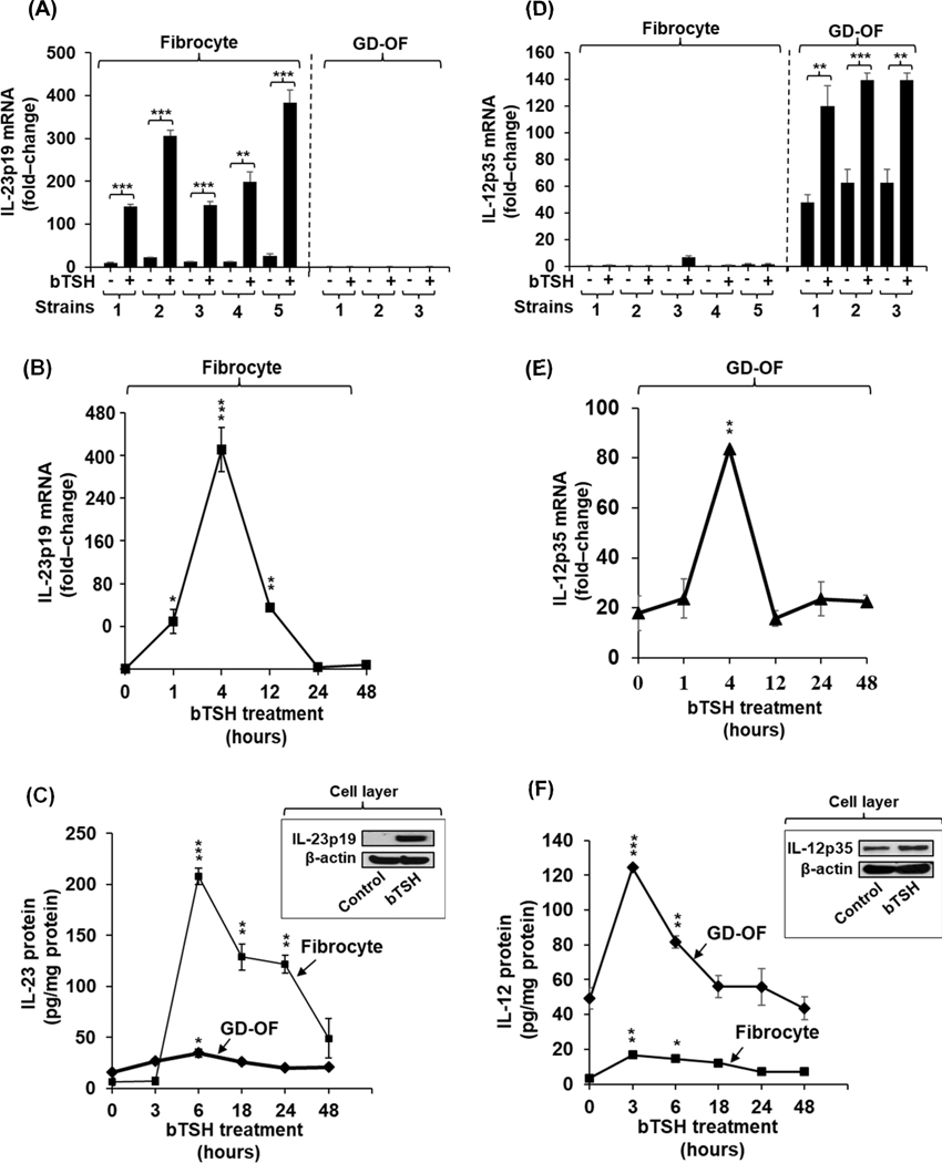
Fig. 1.
Divergent induction by bTSH of IL-23 and IL-12 in GD-fibrocytes versus GD-OF. (A,D) Expression levels of IL-23p19 and IL-12p35 mRNA under control (untreated) and bTSH-treated conditions in 5 strains of GD-fibrocytes and 3 strains of GD-OF, each from a different donor with GD. Medium covering confluent culture monolayers was replaced with DMEM supplemented with 1% FBS and containing nothing or bTSH (5 mIU/mL) for 6 h. Cell layers were harvested, cellular RNA was reverse transcribed, and subjected to quantitative real-time PCR for IL-23p19 and IL-12p35 mRNA. Values were normalized to their respective GAPDH levels and are expressed as the mean ± S.D of triplicate determinations from one representative experiment of three performed, **, p<0.01; ***, p<0.001 by ANOVA. (B and E) Fibrocyte cultures were treated with bTSH (5 mIU/mL) for the graded intervals indicated along the abscissa. IL-23p19 and IL-12p35 mRNA levels were quantified as in A and D and are expressed as mean ± SD. (C and F) Cultures were treated with bTSH for the graded intervals indicated. Media were collected and subjected to IL-23 and IL-12 ELISAs as described in “Methods”. Values were normalized to respective monolayer protein content. Data are expressed as mean ± SD of triplicate determinations. (Insets, C and F) Monolayer proteins from control and bTSH-treated cultures were subjected to Western blot analysis for IL-23p19 and IL-12p35 Blots were then re-probed with anti-β-actin.

