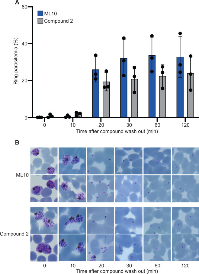
Fig 5. Synchronicity of invasion after removal of inhibition by ML10 or Compound 2.
(A) Parasites that had been arrested in RPMI-1640 supplemented with AlbuMax containing either 25 nM ML10 (blue bars) or 1 μM Compound 2 (grey bars) for up to 3 hours were washed to allow egress and invasion of new erythrocytes. Samples were removed at 10-minute intervals and the percentage of host cells infected with ring forms, which represent parasites that have egressed and invaded new erythrocytes, was determined by flow cytometry (see S3 Fig for gating strategy). Error bars represent standard deviation from three independent experiments each conducted in duplicate, no statistically significant differences were detected between compounds at each time point using a pairwise individual t-test. (B) Giemsa smears of parasites taken at the time points indicated at the top show the stages of egress and invasion, and confirm the presence of ring-stage parasites.

