Figure 1.
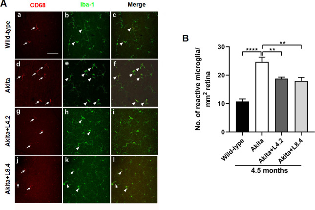
Suppression of microglial reactivity by lutein treatment in the retinas of the Ins2Akita/+ mice. (A) Representative images of the retinal flat-mounts immunostained with CD68 and Iba-1 from the wild-type (a–c), untreated Ins2Akita/+ (d–f), lutein-treated Ins2Akita/+ (4.2 mg/kg/day, Akita+L4.2) (g–i) and lutein-treated Ins2Akita/+ (8.4 mg/kg/day, Akita+L8.4) (j–l) mice at 4.5 months of age. Photomicrographs showed CD68-positive reactive microglia (red, white arrows) and Iba-1-positive total microglia (green, white arrow heads), respectively, together with merged images. Scale bar=100 µm. (B) Quantitative results of number of reactive microglia in the four experimental groups. n=6. Data are presented as mean±SEM; One-way ANOVA followed by Tukey’s multiple comparison test. **P<0.01, ****p<0.0001. ANOVA, analysis of variance; CD68, cluster of differentiation 68; Iba-1, ionized calcium-binding adapter molecule 1.

