
Figure 3.
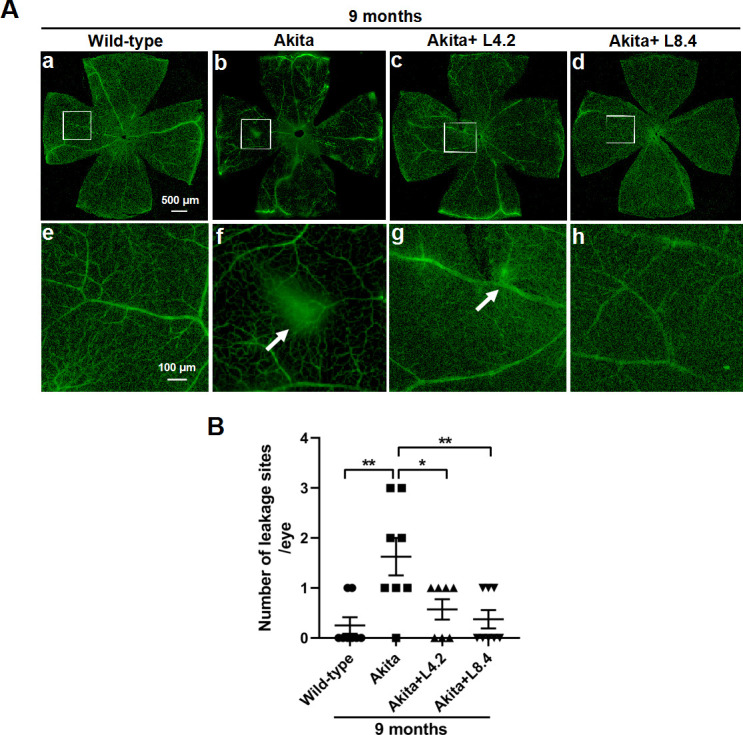
Reduction of retinal vascular leakage by lutein treatment in the retinas of Ins2Akita/+ mice. (A) Images of FITC-dextran-perfused retinal flat-mounts from the wild-type, untreated Ins2Akita/+, lutein-treated Ins2Akita/+ (4.2 mg/kg/day, Akita+L4.2) and lutein-treated Ins2Akita/+ (8.4 mg/kg/day, Akita+L8.4) mice at 9 months of age. Representative images of the entire retinal flat-mounts from the four groups are shown (a–d). Images taken from the corresponding regions in a–d (white rectangles) at higher magnification are shown in e–f. Extravasation of FITC-dextran resulting from retinal vascular damage is shown (white arrows). Scale bars: a–d=500 µm, e–h=100 µm. (B) Quantitative results of number of leakage sites in retinas from four groups. n=7–8. Data are presented as mean±SEM; One-way ANOVA followed by Tukey’s multiple comparison test. *P<0.05, **p<0.01. ANOVA, analysis of variance; FITC, fluorescein isothiocyanate.