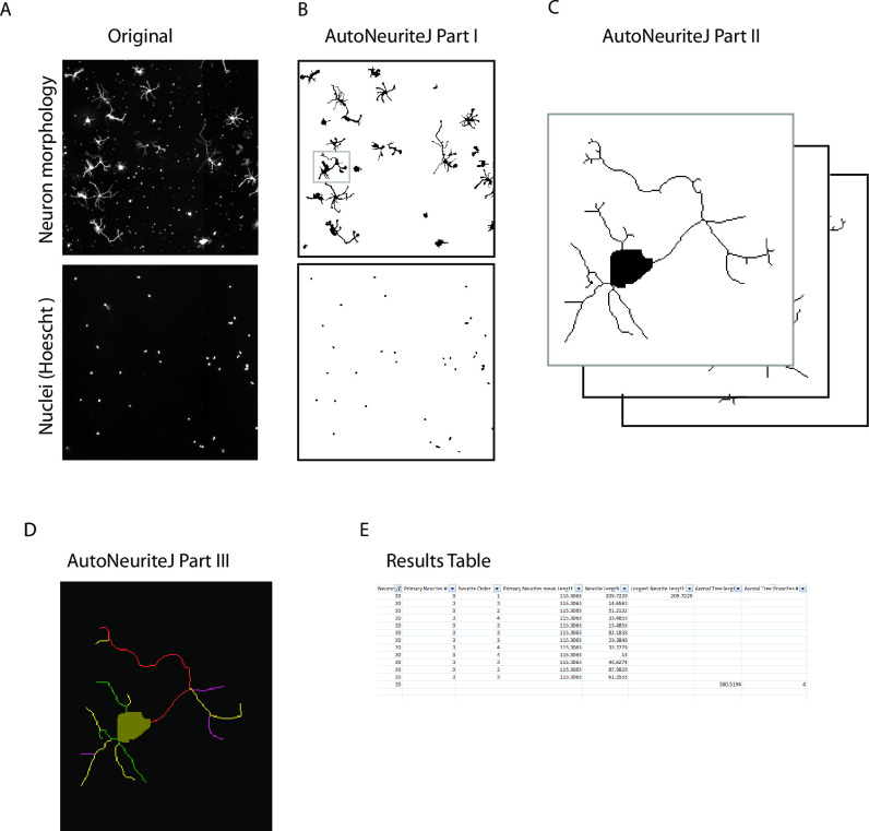
Fig 2. Schematic of AutoNeuriteJ images processing.
(A) AutoNeuriteJ needs images of neuron (e.g. tubulin) and nuclei staining. (B) AutoNeuriteJ Part I segments nuclei and neurons, removes small particles. (C) AutoNeuriteJ part II selects individual neurons (with a single nucleus), creates cell body images and stacks of neurons. (D) AutoNeuriteJ part III detects neurite extremities, compares, classifies and measures neurites, prints results in a text file.

