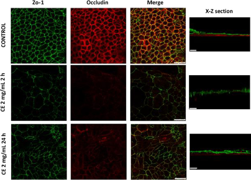
Fig 8. Effects of A. simplex CE on TJ proteins.

Caco-2 cells were incubated with medium or 2mg/mL CE and, thereafter, immunofluorescence staining was performed to localize ZO-1 (green) and occludin (red). TJ proteins distribution is shown at the time of maximal decrease of TEER (2 hours) and recovery of TEER (24 hours). Scale bar represents 50 μm (x-y section) or 20 μm (x-z section).
