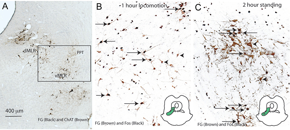
Figure 2. Just medial to the PPN is a population of glutamatergic neurons that project to the spinal cord and are activated during locomotion and standing.
In panel A, the PPN and LDT neurons are stained brown for ChAT, and spinally projecting neurons are immunostained black. In panels B and C, the spinally projecting neurons are labeled brown, and show cFos immunoreactivity (black) in their nucleus if they are activated during forced locomotion (panel B) or forces standing (panel C). Insets show that the locomotion-related neurons are more dorsal than those related to maintaining upright posture. From (Sherman et al., 2015), with permission.

