
Figure 2.
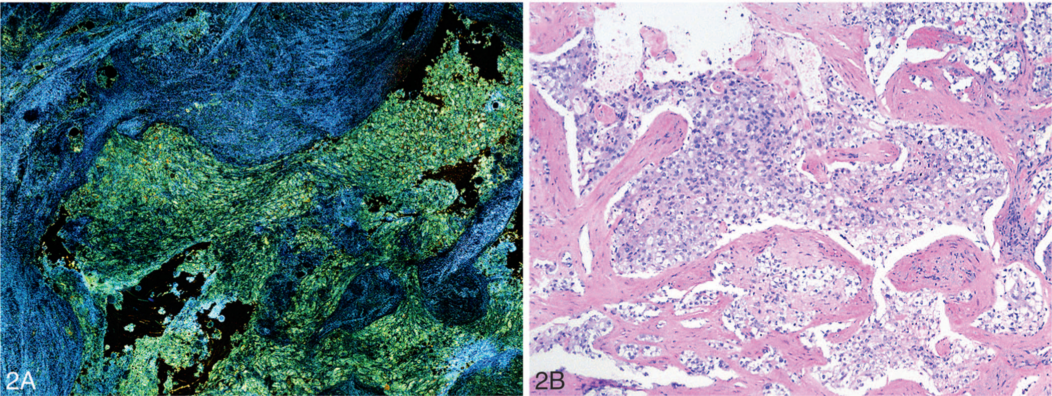
Dynamic full-field optical coherence tomography image (A) and light microscopic image of the corresponding hematoxylin-eosin–stained tissue section (B) of invasive ductal carcinoma of the breast (original magnification ×100 [B]).
Official websites use .gov
A
.gov website belongs to an official
government organization in the United States.
Secure .gov websites use HTTPS
A lock (
) or https:// means you've safely
connected to the .gov website. Share sensitive
information only on official, secure websites.
Dynamic full-field optical coherence tomography image (A) and light microscopic image of the corresponding hematoxylin-eosin–stained tissue section (B) of invasive ductal carcinoma of the breast (original magnification ×100 [B]).